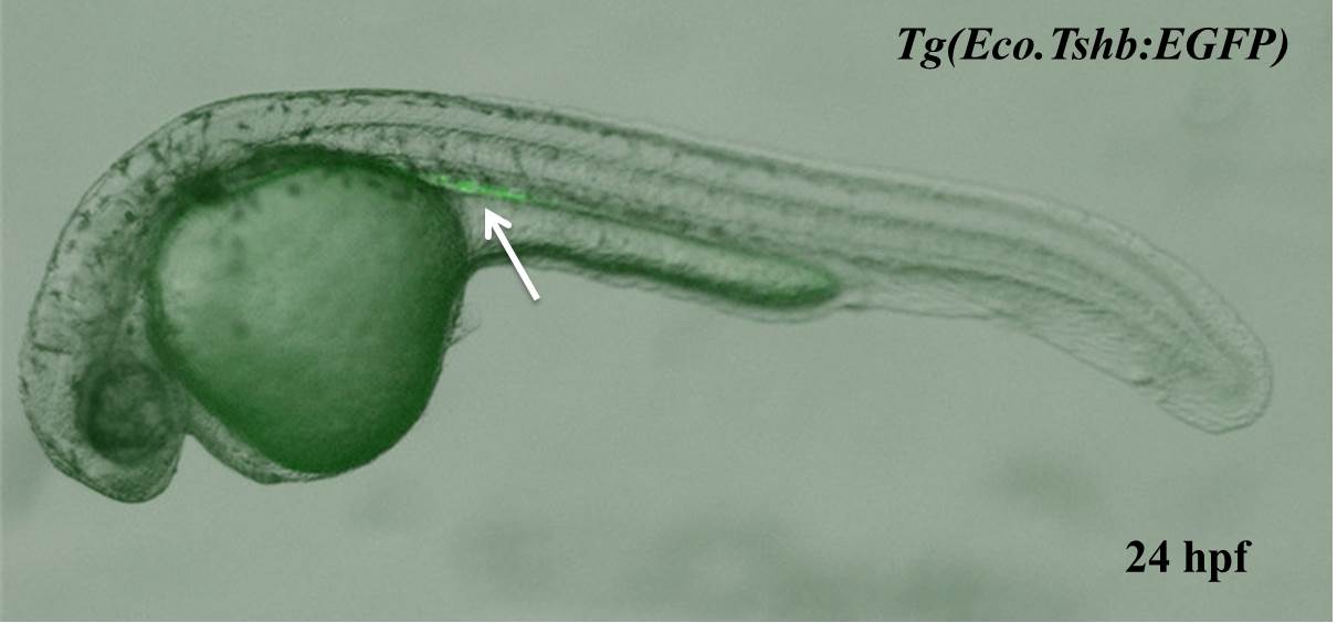
the pronephric neck and tubule | : | the GFP fluorescence signal is initially detectable from 18hpf above the dorsal mesentery and persists throughout the subsequent embryogenesis in Tg(gtshβ:GFP). The initial GFP signal localizes above the anterior region of the yolk extension, and extends from the adjacent fifth somite to the eighth somite. As embryos develop, the GFP fluorescence becomes stronger and extends above the yolk sac, manifesting a bilateral tube-like structure. At 48 hpf, it displays a slight curve, which is likely the pronephric neck and tubule.
|